Microplastics have become a major health concern because of its potential adverse influences on marine, wildlife and public health. In this study, pristine polystyrene and Styrofoam microplastics particles of diameter < 5mm were used to investigate the toxic effects of polystyrene microplastic (PS-MP) exposure on hepatic and renal function of male and female Wistar rats. The rats were divided into seven groups for both male and female, with one control group and six test groups each. The two forms of polystyrene microplastics were incorporated into the feed of the test groups in varying quantities (1, 5 and 10 % of the feed), and exposure lasted for a period of 90 days. Results showed that aspartate aminotransferase (AST), serum albumin (ALB), total bilirubin (TB), conjugated bilirubin (CB) were significantly (p < 0.05) decreased compared to control male and female rats. Histological analysis provided further insights, indicating that despite mild alterations in liver enzymes, albumin and total protein levels in specific test groups, microplastics did not compromise the structural integrity of hepatocytes in male and female rats. However, kidney function parameters exhibited significant (p < 0.05) increases in serum urea, creatinine, K+, and Cl- levels in test rats of both sexes compared to controls. Regardless of sex, the trends of elevated renal markers were similar. These findings suggest that exposure to polystyrene microplastics may adversely affects renal functional capacity even at low doses.
| Published in | Journal of Health and Environmental Research (Volume 10, Issue 2) |
| DOI | 10.11648/j.jher.20241002.12 |
| Page(s) | 41-51 |
| Creative Commons |
This is an Open Access article, distributed under the terms of the Creative Commons Attribution 4.0 International License (http://creativecommons.org/licenses/by/4.0/), which permits unrestricted use, distribution and reproduction in any medium or format, provided the original work is properly cited. |
| Copyright |
Copyright © The Author(s), 2024. Published by Science Publishing Group |
Kidney, Liver, Microplastics, Polystyrene, Styrofoam
PS-MP | Polystyrene Microplastics |
PSP | Polystyrene Pellets |
FP | Processed Polystyrene Plates |
TP | Total Protein |
AST | Aspartate Aminotransferase |
ALB | Albumin |
TB | Total Bilirubin |
CB | Conjugated Bilirubin |
ALT | Alanine Aminotransferase |
ALP | Alkaline Phosphatase |
GGT | Gamma Glutamyl Transferase |
| [1] | Rochman, C. M. (2018). Microplastics research—from sink to source. Science, 360, 28-29. |
| [2] | Frias, J., & Nash, R. (2019). Microplastics: Finding a consensus on the definition. Marine Pollution Bulletin, 138, 145-147. |
| [3] | Rochman, C. M., Browne, M. A., Underwood, A. J., Van-Franeker, J. A., Thompson, R. C., & Amaral-Zettler, L. A. (2016). The ecological impacts of marine debris: Unravelling the demonstrated evidence from what is perceived. Ecology, 97(2), 302-312. |
| [4] | Lu, Y., Zhang, Y., Deng, Y., Jiang, W., Zhao, Y., Geng, J., et al. (2016). Uptake and accumulation of polystyrene microplastics in zebrafish (Danio rerio) and toxic effects in liver. Environmental Science & Technology, 50(7), 4054-4060. |
| [5] | Brun, N. R., Koch, B. E., Varela, M., Peijnenburg, W. J., Spaink, H. P., & Vijver, M. G. (2018). Nanoparticles induce dermal and intestinal innate immune system responses in zebrafish embryos. Environmental Science: Nano, 5, 904-916. |
| [6] | Kögel, T., Bjorøy, Ø., Toto, B., Bienfait, A. M., & Sanden, M. (2020). Micro- and nanoplastic toxicity on aquatic life: Determining factors. Science of the Total Environment, 709, 136050. |
| [7] | Ma, H., Pu, S., Liu, S., Bai, Y., Mandal, S., & Xing, B. (2020). Microplastics in aquatic environments: Toxicity to trigger ecological consequences. Environmental Pollution. Advance online publication. |
| [8] | da Costa, J. P., Santos, P. S., Duarte, A. C., & Rocha-Santos, T. (2016). (Nano) plastics in the environment–sources, fates and effects. Science of the Total Environment, 566, 15-26. |
| [9] | Watts, A. J., Urbina, M. A., Goodhead, R., Moger, J., Lewis, C., & Galloway, T. S. (2016). Effect of microplastic on the gills of the shore crab Carcinus maenas. Environmental Science & Technology, 50(10), 5364-5369. |
| [10] | Horton, A. A., Walton, A., Spurgeon, D. J., Lahive, E., & Svendsen, C. (2017). Microplastics in freshwater and terrestrial environments: Evaluating the current understanding to identify the knowledge gaps and future research priorities. Science of the Total Environment, 586, 127-141. |
| [11] | Lwanga, E. H., Gertsen, H., Gooren, H., Peters, P., Salánki, T., & van der Ploeg, M., et al. (2017). Incorporation of microplastics from litter into burrows of Lumbricus terrestris. Environmental Pollution, 220, 523-531. |
| [12] | Rist, S., Almroth, B. C., Hartmann, N. B., & Karlsson, T. M. (2018). A critical perspective on early communications concerning human health aspects of microplastics. Science of the Total Environment, 626, 720-726. |
| [13] | Irving, R. M., & Elfarra, A. A. (2012). Role of reactive metabolites in the circulation in extrahepatic toxicity. Expert Opinion on Drug Metabolism & Toxicology, 8(9), 1157-1172. |
| [14] | Mahadappa, P., Krishnaswamy, N., Karunanidhi, M., Bhanuprakash, A. G., Bindhuja, B. V., & Dey, S. (2020). Effect of plastic foreign body impaction on rumen function and heavy metal concentrations in various body fluids and tissues of buffaloes. Ecotoxicology and Environmental Safety, 189, 109972. |
| [15] | Rochman, C. M. (2013). Plastics and priority pollutants: A multiple stressor in aquatic habitats. Environmental Science & Technology, 47(5), 2439–2440. |
| [16] | Ogbu, S. I., & Okechukwu, F. I. (2001). The effect of storage temperature prior to separation on plasma and serum potassium. Journal of Medical Laboratory Science, 10, 1-4. |
| [17] | Doumas, B. T., Watson, W. A., & Biggs, H. G. (1971). Albumin standards and measurement of serum-albumin with bromocresol green. Clinical Chimica Acta, 31, 87. |
| [18] | Evelyn, K. A., & Malloy, H. T. (1938). Micro determination of oxyhaemoglobin, methaemoglobin and sulphaemoglobin in a single sample of blood. Journal of Biological Chemistry, 126, 655. |
| [19] | Tietz, N. W., Prude, E. L., & Sirgard-Anderson, O. (1994). Tietz Textbook of Clinical Chemistry (2nd ed.). W. B. Saunders Company. |
| [20] | Kaplan, A. (1965). Urea Nitrogen and Urinary Ammonia. In S. Meites (Ed.), Standard Method of Clinical Chemistry (pp. 245-256). Academic Press Inc. |
| [21] | Bassir, O. (1971). Handbook of Practical Biochemistry. Ibadan University Press. |
| [22] | Revel, M., Chatel, A., & Mouneyrac, C. (2018). Micro (nano) plastics: A threat to human health? Current Opinion in Environmental Science & Health, 1, 17-23. |
| [23] | Jeong, C. B., Kang, H. M., Lee, M. C., et al. (2017). Adverse effects of microplastics and oxidative stress-induced MAPK/Nrf2 pathway-mediated defense mechanisms in the marine copepod Paracyclopina nana. Scientific Reports, 7, 41323. |
| [24] | Deng, Y., Zhang, Y., Lemos, B., & Ren, H. (2017). Tissue accumulation of microplastics in mice and biomarker responses suggest widespread health risks of exposure. Scientific Reports, 7, 46687. |
| [25] | von Moos, N., Burkhardt-Holm, P., & Köhler, A. (2012). Uptake and effects of microplastics on cells and tissue of the blue mussel Mytilus edulis L. after an experimental exposure. Environmental Science & Technology, 46(20), 11327-11335. |
| [26] | Obi, C. L., Bessong, P. O., Momba, M. N. B., Potgieter, N., Samie, A., & Igumbor, E. O. (2004). Profiles of antibiotic susceptibilities of bacterial isolates and physico-chemical quality of water supply in rural Venda communities, South Africa. Water SA, 30(4), 515-519. |
| [27] | Bhuyan, M. S. (2022). Effects of Microplastics on Fish and in Human Health. Frontiers in Environmental Science, 10, 827289. |
| [28] | Okolie, E. C. (2011). Geoelectric investigation of the effect of heavy clay deposits on aquifer potential in Okpara waterside Delta State, Nigeria. Journal of Geology and Mining Research, 3(2), 39-45. |
| [29] | Agbasi, K. C., Ezeani, M. I., Osita, E. G., Okonkwo, R. O., & Nnanwa, C. P. (2010). Heuristic Approaches to Solving MultiObjective Scheduling Optimisation Problems. Electroscope Journal, 4(4), 1-12. |
| [30] | Ogunka-Nnoka, C., Amadi, B., Agomuo, E., & Amadi, P. (2017). Ameliorative effects of some natural blood boosters on cyclophosphamide-induced anaemia in rats. Journal of Applied Life Sciences International, 10(3), 1-11. |
| [31] | Yang, Y. F., Chen, C. Y., Lu, T. H., & Liao, C. M. (2019). Toxicity-based toxicokinetic/toxicodynamic assessment for bioaccumulation of polystyrene microplastics in mice. Journal of Hazardous Materials, 366, 703–713. |
| [32] | Wang, Y., Lee, Y., Hsu, Y., Chiu, I., Huang, C. C., Huang, C., Chia, Z., Lee, C., Lin, Y., & Chiu, H. (2021). The kidney-related effects of polystyrene microplastics on human kidney proximal tubular epithelial cells HK-2 and male C57BL/6 mice. Environmental Health Perspectives, 129(5), 1-18. |
| [33] | Chatterjea, M. N., & Sinde, R. (2012). Metabolism of Minerals and Trace Elements. In M. N. Chatterjea & R. Sinde (Eds.), Textbook of Medical Biochemistry (8th ed., pp. 608-628). Jaypee Brothers Medical Publishers. |
| [34] | Kang, I. S., Jin, K., Wang, B., Lau, K. M., Shukla, J., Krishnamurthy, V., & Liu, Y. (2002). Intercomparison of the climatological variations of Asian summer monsoon precipitation simulated by 10 GCMs. Climate Dynamics, 19(5), 383-395. |
| [35] | Nduka, N. (1999). Water and electrolytes. In N. Nduka (Ed.), Clinical Biochemistry for Students of Pathology. Longman Nigeria Plc. Pp. 23–30. |
APA Style
Okonkwo, C. J., Nnoruka, U. C., Okonkwo, C. J., Ilechukwu, I., Belonwu, D. C. (2024). Impact of Polystyrene Exposure on Hepatorenal Responses in Male and Female Albino Wistar Rats. Journal of Health and Environmental Research, 10(2), 41-51. https://doi.org/10.11648/j.jher.20241002.12
ACS Style
Okonkwo, C. J.; Nnoruka, U. C.; Okonkwo, C. J.; Ilechukwu, I.; Belonwu, D. C. Impact of Polystyrene Exposure on Hepatorenal Responses in Male and Female Albino Wistar Rats. J. Health Environ. Res. 2024, 10(2), 41-51. doi: 10.11648/j.jher.20241002.12
AMA Style
Okonkwo CJ, Nnoruka UC, Okonkwo CJ, Ilechukwu I, Belonwu DC. Impact of Polystyrene Exposure on Hepatorenal Responses in Male and Female Albino Wistar Rats. J Health Environ Res. 2024;10(2):41-51. doi: 10.11648/j.jher.20241002.12
@article{10.11648/j.jher.20241002.12,
  author = {Chinedu Joseph Okonkwo and Udoka Chukwudubem Nnoruka and Chioma Joy Okonkwo and Ifenna Ilechukwu and Donatus Chuka Belonwu},
  title = {Impact of Polystyrene Exposure on Hepatorenal Responses in Male and Female Albino Wistar Rats
},
  journal = {Journal of Health and Environmental Research},
  volume = {10},
  number = {2},
  pages = {41-51},
  doi = {10.11648/j.jher.20241002.12},
  url = {https://doi.org/10.11648/j.jher.20241002.12},
  eprint = {https://article.sciencepublishinggroup.com/pdf/10.11648.j.jher.20241002.12},
  abstract = {Microplastics have become a major health concern because of its potential adverse influences on marine, wildlife and public health. In this study, pristine polystyrene and Styrofoam microplastics particles of diameter < 5mm were used to investigate the toxic effects of polystyrene microplastic (PS-MP) exposure on hepatic and renal function of male and female Wistar rats. The rats were divided into seven groups for both male and female, with one control group and six test groups each. The two forms of polystyrene microplastics were incorporated into the feed of the test groups in varying quantities (1, 5 and 10 % of the feed), and exposure lasted for a period of 90 days. Results showed that aspartate aminotransferase (AST), serum albumin (ALB), total bilirubin (TB), conjugated bilirubin (CB) were significantly (p < 0.05) decreased compared to control male and female rats. Histological analysis provided further insights, indicating that despite mild alterations in liver enzymes, albumin and total protein levels in specific test groups, microplastics did not compromise the structural integrity of hepatocytes in male and female rats. However, kidney function parameters exhibited significant (p < 0.05) increases in serum urea, creatinine, K+, and Cl- levels in test rats of both sexes compared to controls. Regardless of sex, the trends of elevated renal markers were similar. These findings suggest that exposure to polystyrene microplastics may adversely affects renal functional capacity even at low doses.
},
 year = {2024}
}
TY  - JOUR
T1  - Impact of Polystyrene Exposure on Hepatorenal Responses in Male and Female Albino Wistar Rats

AU  - Chinedu Joseph Okonkwo
AU  - Udoka Chukwudubem Nnoruka
AU  - Chioma Joy Okonkwo
AU  - Ifenna Ilechukwu
AU  - Donatus Chuka Belonwu
Y1  - 2024/07/31
PY  - 2024
N1  - https://doi.org/10.11648/j.jher.20241002.12
DO  - 10.11648/j.jher.20241002.12
T2  - Journal of Health and Environmental Research
JF  - Journal of Health and Environmental Research
JO  - Journal of Health and Environmental Research
SP  - 41
EP  - 51
PB  - Science Publishing Group
SN  - 2472-3592
UR  - https://doi.org/10.11648/j.jher.20241002.12
AB  - Microplastics have become a major health concern because of its potential adverse influences on marine, wildlife and public health. In this study, pristine polystyrene and Styrofoam microplastics particles of diameter < 5mm were used to investigate the toxic effects of polystyrene microplastic (PS-MP) exposure on hepatic and renal function of male and female Wistar rats. The rats were divided into seven groups for both male and female, with one control group and six test groups each. The two forms of polystyrene microplastics were incorporated into the feed of the test groups in varying quantities (1, 5 and 10 % of the feed), and exposure lasted for a period of 90 days. Results showed that aspartate aminotransferase (AST), serum albumin (ALB), total bilirubin (TB), conjugated bilirubin (CB) were significantly (p < 0.05) decreased compared to control male and female rats. Histological analysis provided further insights, indicating that despite mild alterations in liver enzymes, albumin and total protein levels in specific test groups, microplastics did not compromise the structural integrity of hepatocytes in male and female rats. However, kidney function parameters exhibited significant (p < 0.05) increases in serum urea, creatinine, K+, and Cl- levels in test rats of both sexes compared to controls. Regardless of sex, the trends of elevated renal markers were similar. These findings suggest that exposure to polystyrene microplastics may adversely affects renal functional capacity even at low doses.

VL  - 10
IS  - 2
ER  -
Department of Biochemistry, Faculty of Science, University of Port Harcourt, Rivers, Nigeria
Department of Biochemistry, Faculty of Science, University of Port Harcourt, Rivers, Nigeria
Department of Biochemistry, Faculty of Science, University of Port Harcourt, Rivers, Nigeria
Graduate School of Engineering and Science, Department of Marine and Environmental Science, University of the Ryukyus, Nishihara, Japan; Environmental Chemistry Unit, Department of Industrial Chemistry, Madonna University, Elele Campus, Rivers, Nigeria
Department of Biochemistry, Faculty of Science, University of Port Harcourt, Rivers, Nigeria
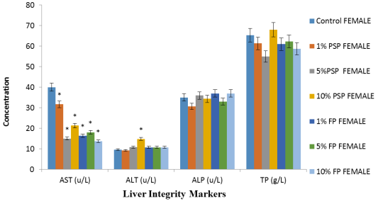
Figure 1. Effects of polystyrene exposure on serum aspartate aminotransferase (AST), alanine aminotransferase (ALT), alkaline phosphatase (ALP) and total protein (TP) of Female Wistar rats.
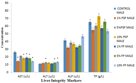
Figure 2. Effects of polystyrene exposure on serum aspartate aminotransferase (AST), alanine aminotransferase (ALT), alkaline phosphatase (ALP) and total protein (TP) of Male Wistar rats.
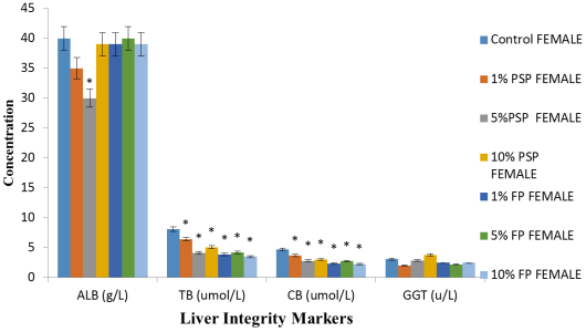
Figure 3. Effects of polystyrene exposure on serum serum albumin (ALB), total bilirubin (TB), conjugated bilirubin (CB) and gamma-glutamyl transpeptidase (GGT) of Female Wistar rats.
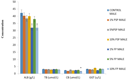
Figure 4. Effects of polystyrene exposure on serum serum albumin (ALB), total bilirubin (TB), conjugated bilirubin (CB) and gamma-glutamyl transpeptidase (GGT) of Female Wistar rats.
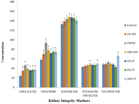
Figure 5. Concentrations of serum urea, creatinine and electrolytes of female Wistar rat exposed to different percentage of polystyrene MP compared to the control.
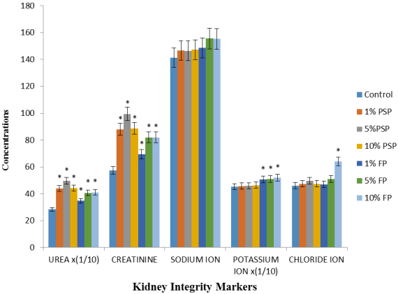
Figure 6. Concentrations of serum urea, creatinine and electrolytes of male Wistar rat exposed to different percentage of polystyrene MP compared to the control.
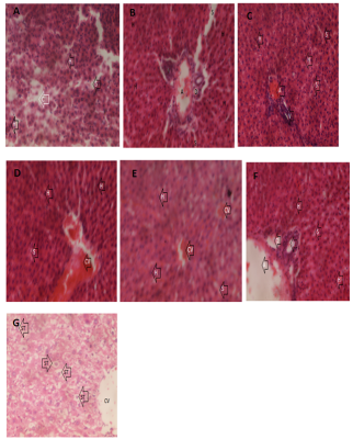
Figure 7. (A-G) Histological assessment of female rat liver sections stained with hematoxylin and eosin (x400). (A) Group 1 (Control): showing no lesions or abnormality; (B) Group 2 (1% PSP): showing normal hepatocytes; (C) Group 3 (5% PSP): showing intact hepatocytes (H) and sinusoids (S); (D) Group 4 (10% PSP): showing congested central vein, intact hepatocytes (H) and sinusoids (S); (E) Group 5 (1% FP): showing intact hepatocytes (H) and sinusoids (S); (F) Group 6 (5% FP): showing intact hepatocytes (H) and sinusoids (S); (G) Group 7 (10% PSP): showing intact hepatocytes (H) radiating away from central vein (CV) and sinusoids (S).
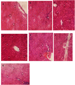
Figure 8. (A-G) Histological assessment of male rat liver sections stained with hematoxylin and eosin (x400). (A) Group 1 (Control): showing histologically normal liver with intact hepatocytes; (B) Group 2 (1% PSP): showing mildly distorted liver with microvesicular steatosis; (C) Group 3 (5% PSP): showing intact hepatocytes (H) and sinusoids (S); (D) Group 4 (10% PSP): showing patent central vein, intact hepatocytes (H) and sinusoids (S); (E) Group 5 (1% FP): showing intact hepatocytes (H) and sinusoids (S); (F) Group 6 (5% FP): showing congested central vein, intact hepatocytes (H) and sinusoids (S); (G) Group 7 (10% PSP): showing intact hepatocytes (H), sinusoids (S) and portal triad (hepatic artery (A), Portal vein (V) and bile duct (D)).
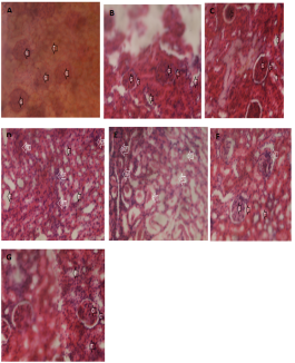
Figure 9. (A-G) Histological assessment of female rat kidney sections stained with hematoxylin and eosin (x400). (A) Group 1 (Control): showing no lesions or abnormality; (B) Group 2 (1% PSP): showing normal kidney with intact Glomeruli (G) and patent Bowman’s capsule (C); (C) Group 3 (5% PSP): showing intact kidney glomerulus (G) and Bowman’s capsule (C); (D) Group 4 (10% PSP): showing distorted kidney showing interstitial tissues filled with inflammatory cells (INF); (E) Group 5 (1% FP): showing distorted kidney with damaged renal tubule (DT); (F) Group 6 (5% FP): showing distorted kidney with interstitial tissues filled with inflammatory cells; (G) Group 7 (10% PSP): showing distorted kidney.
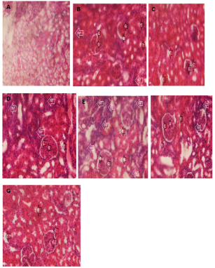
Figure 10. (A-G) Histological assessment of male rat kidney sections stained with hematoxylin and eosin (x400). (A) Group 1 (Control): showing no lesions or abnormality; (B) Group 2 (1% PSP): showing normal kidney with intact Glomeruli (G) and patent Bowman’s capsule (C); (C) Group 3 (5% PSP): showing normal kidney glomerulus (G) and Bowman’s capsule (C); (D) Group 4 (10% PSP): showing distorted kidney showing interstitial tissues filled with inflammatory cells (INF); (E) Group 5 (1% FP): showing kidney with interstitial tissues filled with inflammatory cells (INF); (F) Group 6 (5% FP): showing distorted kidney with interstitial tissues filled with inflammatory cells; (G) Group 7 (10% PSP): showing distorted kidney.